


Summary
The retina is the light-detecting tissue located at the back of the eye. At its centre lies the macula, the area responsible for detailed and focused vision. A macular hole is a tiny break that forms in this central region. Although small in size, it can greatly disturb everyday activities such as reading, identifying faces, or driving.
The eye is an extremely sensitive organ, and even minor structural changes can affect eyesight. A macular hole occurs when a small full-thickness gap develops in the centre of the retina. Age is the most common reason behind this condition, especially in individuals above 55–60 years. While the opening may appear insignificant, its effect on central vision can be serious.
This condition is relatively uncommon and affects a limited number of people. In rural regions of Bihar and Uttar Pradesh, awareness about retinal disorders remains low, which often delays proper treatment. Learning about macular holes can help individuals notice early warning signs and seek timely medical care.
What Is a Macular Hole?
The retina forms the inner lining at the back of the eye and helps convert light into visual signals. At its centre is the macula, a small but highly important area that enables us to see fine details clearly. Tasks such as reading small print, recognising faces, threading a needle, or driving depend heavily on the macula.
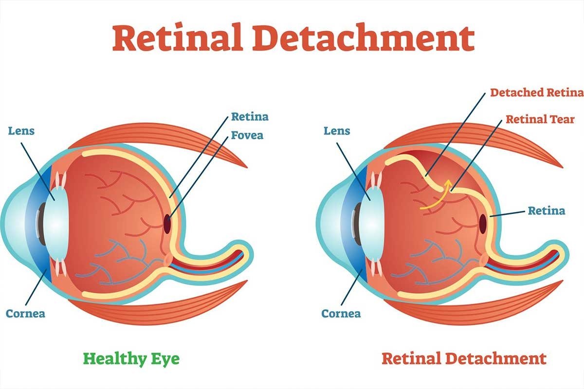
A macular hole develops when a small opening forms in this central region. It is different from a retinal tear because it specifically involves the macula rather than the peripheral retina. Even though the defect is tiny, its location makes it highly impactful on vision.
People sometimes mistake it for age-related macular degeneration or retinal detachment because the symptoms may overlap. However, a macular hole is a distinct medical problem with separate causes and management.
How Does a Macular Hole Form?
With ageing, natural changes occur inside the eye. The vitreous — a gel-like substance that fills the eye — gradually shrinks over time. Normally, it separates gently from the retina without causing harm. In some cases, however, the vitreous remains partially attached to the macula and pulls on it. This traction can stretch the tissue and eventually create a hole.
Although ageing is the primary cause, other contributing factors include:
1. Eye Trauma
A strong injury can disturb the delicate retinal structure. Sudden force may create traction on the macula and lead to a hole.
2. High Myopia (Severe Near-Sightedness)
Individuals with significant myopia often have thinner retinal tissue. This structural weakness increases the chances of developing macular damage.
3. Chronic Macular Swelling
Long-term inflammation or fluid accumulation in the macula can gradually weaken the tissue, making it more prone to forming a hole.
4. Previous Eye Surgery
In rare cases, internal changes following certain eye surgeries may contribute to the development of a macular hole.
Who Is More Likely to Develop It?
Although anyone can develop this condition, certain groups are at higher risk:
- Adults over 50 years of age
- People with high myopia
- Individuals who previously had a macular hole in one eye
- Those with a history of eye injury or chronic eye inflammation
With an increasing elderly population and many untreated refractive errors in Bihar, recognising these risk factors can encourage earlier diagnosis and treatment.
Symptoms of a Macular Hole
Because the macula controls central vision, symptoms directly affect focused sight. Common signs include:
- A blurred or dark spot in the centre of vision
- Straight lines appearing bent or distorted
- Difficulty reading even large text
- Gradual worsening of clarity
Vision loss usually progresses slowly, though it may sometimes develop suddenly. While total blindness does not occur, central vision impairment can significantly interfere with daily life.
Stages of a Macular Hole
Doctors classify macular holes into stages to determine severity and treatment planning.
Stage 1 (Foveal Detachment):
The macula begins to stretch, but a complete hole has not yet formed. Early treatment at this stage may prevent progression.
Stage 2:
A small partial-thickness opening develops. Visual distortion becomes more noticeable.
Stage 3:
The hole extends through the full thickness of the macula, leading to marked central vision loss.
Stage 4:
The vitreous completely separates from the retina, and the hole enlarges further. Daily tasks become extremely challenging at this stage.
Early detection plays a key role in better visual recovery.
Diagnosis
An eye specialist performs a detailed retinal examination to confirm the condition. The most advanced and reliable test is an OCT (Optical Coherence Tomography) scan. This painless imaging technique provides cross-sectional views of the retina, allowing doctors to detect even very small macular openings.
Treatment Options
In most cases, a macular hole does not close on its own. Medicines such as eye drops are usually ineffective. The most dependable treatment is a surgical procedure known as vitrectomy.
Vitrectomy Surgery
During this operation, the surgeon removes the vitreous gel that is pulling on the macula. A special gas bubble is then placed inside the eye. This bubble gently presses against the macula, helping the hole to seal gradually.
Conclusion
A macular hole is a small defect in the central retina, but its impact on detailed vision can be considerable. It mainly affects older adults, and awareness remains limited in many rural areas of Bihar. Understanding its causes, recognising early symptoms, and seeking timely surgical care can help prevent permanent central vision loss.
Refresh Date: March 3, 2026